


Shaping Tomorrow's Hip Replacement
DU’s Center for Orthopaedic Biomechanics studies pelvic mobility to improve patient outcomes.
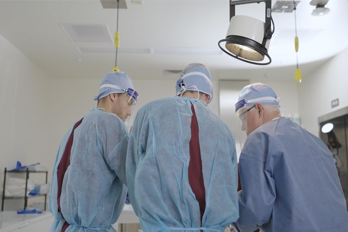
Many of us have had or know people who have had hip replacement surgeries, but few of us think about what really happens on the operating table.
For example, did you know that traditional total hip replacement procedures require big incisions through the gluteus muscles that enable surgeons to clearly see the hip joint?
That’s as intrusive as it sounds. But there’s another, less intrusive way to perform the surgery—and a team of researchers in the Daniel Felix Ritchie School of Engineering and Computer Science is improving the odds of its success.

As Chadd Clary, associate professor of mechanical and materials engineering, explains, “Traditional hip replacement is a very intrusive process because you're separating big muscles to get access to the hip joint below.” However, he adds, “In the last decade or so, there's been a big shift to performing total hip replacement through an anterior approach. The incision comes in on the front side of the hip.”
A more direct path to the joint via smaller incisions means the surgeon is cutting through less muscle, which improves patient recovery—but this approach reduces the surgeon’s view of the joint.
Clary and his team are working on giving surgeons the proper tools to get implants into the right orientation when their visibility is reduced. With the support of a grant from Mizuho OSI, a San Francisco Bay-area company that specializes in surgical tables, they are assessing pelvic mobility during anterior-approach total hip replacement using the company’s Hana surgical table.
The Hana table allows for changes to pelvic orientation during surgery. “When you have surgery, your body is manipulated. It's pushed on. It's reoriented. They have to dislocate your hip to place the components,” Clary explains. The table also permits X-rays during operations to confirm implant placement.
The procedure, however, is complicated by the fact that everyone’s pelvis moves differently. “The way your pelvis moves is very much a characteristic of who you are. Everybody's pelvis seems to move in its own way,” explains Casey Myers, research associate professor of mechanical and materials engineering. The Ritchie team is documenting this special movement, which they refer to as a patient-specific approach to pelvic mobility.
Another key factor is that, when surgeons insert an artificial joint into a patient, they’re seeing the orientation of the pelvis on the operating table—which, Clary says, “is not necessarily a reflection of where the pelvis is when someone is standing. When you stand that pelvis can take a very different orientation that is influenced by your lumbar spine.”
Various tests can help surgeons make the proper adjustments, such as fluoroscopy—taking moving X-rays of patients as they perform tasks such as sitting in a chair, standing up and walking. The team correlates the functional orientation of the pelvis during these dynamic activities to their supine position on the Hana table. That helps determine what factors need to be accounted for during surgery.
DU’s Center for Orthopaedic Biomechanics is uniquely equipped to study these factors, with its suite of six laboratories for data collection in areas such as computational biomechanics, human dynamics, and biomaterials and testing.
“DU’s a unique site because the labs complement each other,” says Myers. “We're able to take a question and come at it from different directions to have a comprehensive solution or description of what we're after.”
Team member Kathryn Colone, a third-year PhD student, lauds the array of technology available in the labs, such as computer modeling, laser scanners, 3D printing for special experimental components and equipment that simultaneously collects motion and force data.
“When I chose DU, I realized this is exactly what I’d been looking for. I couldn’t find it anywhere else. I’m very fortunate,” Colone says.
The team is able to work with real patients through partnerships with Dr. Joseph Assini of Swedish Medical Center in Englewood, Colorado, who performs hip replacement surgeries, and DePuy Synthes, a medical device subsidiary of Johnson and Johnson, which is providing funding for “in vivo” data collection—studying how the pelvises of patients move during functional activities in the lab.
“To pull this off, at any point in time, we probably had 10 or 12 people in the room doing different components of making this experiment run the way that it was intended to,” Clary says.
The team excels at problem-solving and thinking collaboratively, Colone says. “No matter what the challenges are during testing, we always find a way to collect the data we need to get the results we’re looking for.”
Colone says she’s planning to continue working in the field of medical devices and orthopaedics after graduation. “Everything I'm learning here is applicable to what I could be doing anywhere outside the lab, with the amount of knowledge I've gained,” she says. “All the different technologies we've used will translate nicely into whatever field I end up doing research.”
Everyone on the team chose the field of orthopaedic biomechanics because they wanted to help improve the quality of people’s lives.
“If you have hip pain, and it's affecting your quality of life, you shouldn't put off doing a hip replacement out of a lack of understanding of what's involved,” Clary says. “I know it can be scary to get surgery, but the outcomes are fantastic.”


